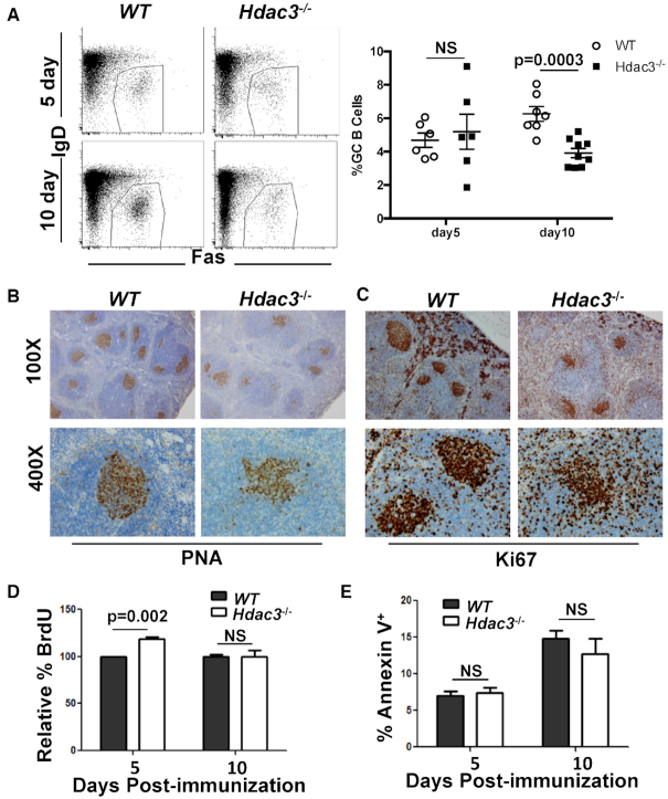
Figure 4.
Altered germinal centers in the absence of Hdac3. (A) Mice were immunized with sheep red blood cells (SRBCs). 5 and 10 days post-immunization, spleens were harvested and analyzed by flow cytometry for the presence of B220+IgDloFas+ germinal center B cells. Graphical representation of GC B cell populations as a percentage of total splenic B cells from at least 6 mice per group is presented at the right. (B) Spleens were isolated from SRBC-injected mice ten days following immunization and sections were stained with PNA to identify germinal center B cells. (C) Ki67 staining was performed on 10 dpi spleens to identify proliferating cells. (D) Immunized mice were injected with BrdU 2 h prior to tissue harvest. Germinal center B cells were analyzed by flow cytometry for BrdU incorporation. (E) Splenocytes were labeled with annexin V and analyzed by flow cytometry. The percentage of B220+IgDloFas+ Annexin V+ splenocytes are depicted.

