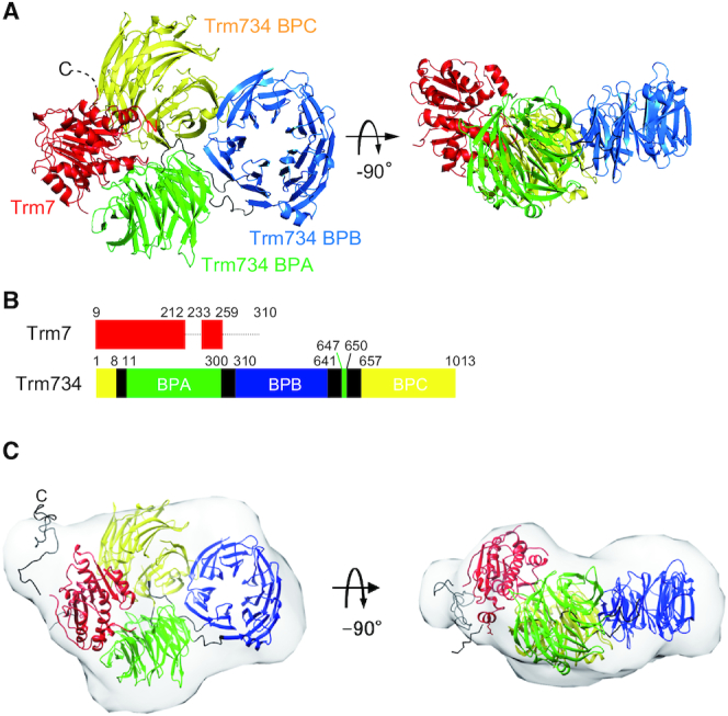
Figure 2.

Structure of Trm7–Trm734. (A) Ribbon diagram of the X-ray structure of Trm7–Trm734. Trm7 is colored red. The three domains of Trm734 (BPA, BPB and BPC) and linker regions are colored green, blue, yellow and black, respectively. The structures on the right are rotated through −90° along the horizontal axis. (B) Schematic representation of Trm7 and Trm734 with domain boundaries. The dotted lines show the regions of Trm7 which are invisible. (C) Overlay of the envelope shape of Trm7–Trm734 calculated with DAMMIN using the SAXS data and the CORAL model of Trm7–Trm734 based on its X-ray structure.
