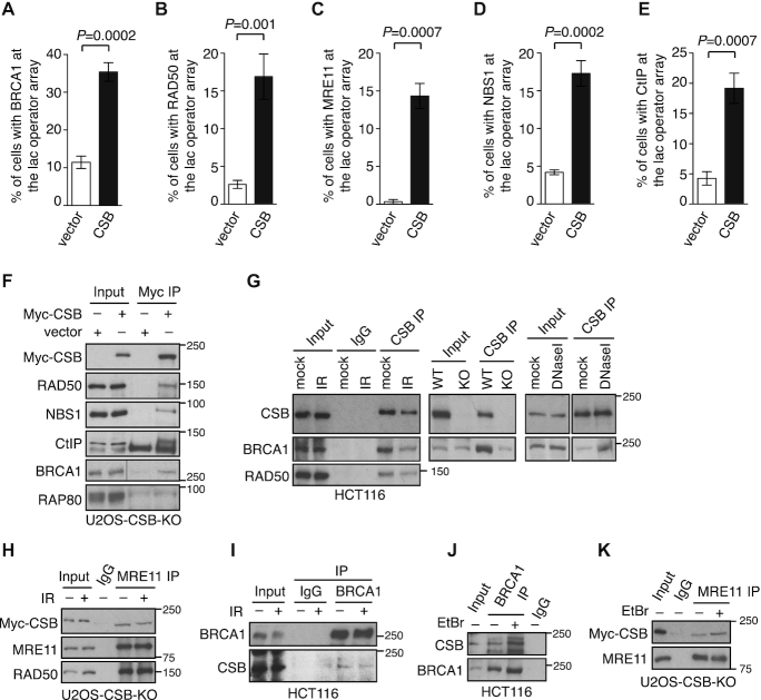
Figure 2.
CSB interacts with the BRCA1-C complex. (A–E) CSB colocalizes with the BRCA1-C complex at the lac operator array. Quantification of vector- and mCherry-LacR-CSB-expressing U2OS-265 CSB-KO cells exhibiting an accumulation of BRCA1 (A), RAD50 (B), MRE11 (C), NBS1 (D), CtIP (E) at the lac operator array. At least 100 cells positive for mCherry staining were scored per condition in a blind manner. Standard deviations from three independent experiments are indicated. (F) CoIPs with anti-Myc antibody in U2OS CSB-KO cells expressing the vector alone or Myc-CSB. Immunoblotting was performed with antibodies against various proteins as indicated in this and subsequent Figures. (G) Anti-CSB coIPs done with HCT116 cells treated with or without 10 Gy IR (the left panel), with HCT116 WT or CSB-KO cells (the middle panel) and with HCT116 cells treated with or without 100 units/ml DNAase I (the right panel). (H) Anti-MRE11 coIPs of Myc-CSB-expressing U2OS CSB-KO cells treated with or without 10 Gy IR. (I) Anti-BRCA1 coIPs of HCT116 cells treated with or without 10 Gy IR. (J) Anti-BRCA1 coIPs of HCT116 cells treated with or without 100 ng/ml ethidium bromide (EtBr). (K) Anti-MRE11 coIPs of Myc-CSB-expressing U2OS CSB-KO cells treated with or without 100 ng/ml EtBr.

