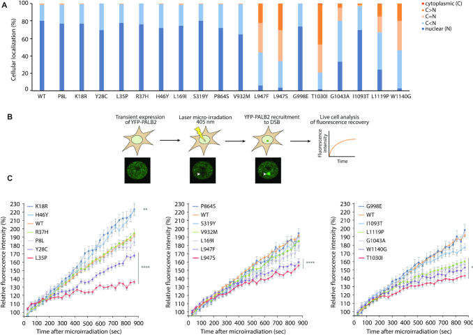
Figure 3.
Subcellular localization and recruitment of PALB2 variants to DNA damage. (A) Subcellular localization of YFP-PALB2 missense variants compared to the WT protein (n > 120 cells per condition). (B) Schematic representation of the laser micro-irradiation experiment used for analyzing the recruitment kinetics of PALB2. (C) Quantitative evaluation of recruitment kinetics for YFP-PALB2 WT or missense variants to laser-induced DSBs. Mean curves ± SEM are shown (n > 60 cells per condition). Statistics were performed on the last time point (900 s time point) using Kruskal–Wallis test followed by Dunn's multiple comparison post-test. Data from A and C are from at least three independent experiments in HeLa cells. (**) P< 0.01 and (****) P< 0.0001.

