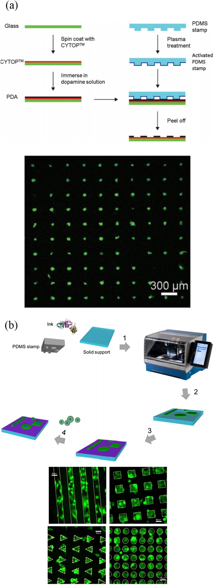
FIG. 9.
High-throughput microcontact printing methods and results. (a) Schematic illustration of the fabrication of PDA patterns on the CYTOP-coated glass surface by negative microcontact printing. Single cell array is formed on the PDA-patterned CYTOP surface. Reproduced with permission from H. Wu, L. Wu, X. Zhou, B. Liu, and B. Zheng, Small 14, e1802128 (2018). Copyright 2018 WileyVCH Verlag GmbH & Co. KGaA, Weinheim. (b) Overall microcontact printing of adhesive patterns using InnoStamp. PC3-GFP cell microarrays are formed on fibronectin micropatterns of various shapes. Reproduced with permission from J. Foncy, A. Estève, A. Degache, C. Colin, J. C. Cau, L. Malaquin, C. Vieu, and E. Trévisiol, Methods Mol. Biol. 1771, 83 (2018). Copyright 2018 Springer Science Business Media, LLC, part of Springer Nature.

