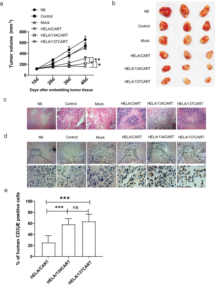
Figure 4.
Antitumor activity of HELA/CART cells in a xenograft model in vivo. (A) BALB/c nude mice were inoculated with SUNE1-LMP1 cells overexpressing LMP1 and allowed to develop established tumors over 10 days. The results are expressed as the mean tumor volume (mm3) (mean±SD) for the six groups (N=6/group). The HELA/137CART group was able to inhibit tumor growth more effectively than the HELA/CART and HELA/134CART groups. *p<0.05; **p<0.01. (B) Images of representative tumor samples from sacrificed mice are shown. (C) The tumor samples were paraffin-embedded and stained with H&E. The tumor tissue necrosis area of the HELA/CART, HELA/134CART, and HELA/137CART treatment groups was larger and showed more lymphocyte infiltration, compared with control groups. (D) Immunohistochemical (IHC) staining for anti-human CD3ζ was performed on tumor samples. HELA/134CART and HELA/137CART cells were more likely to infiltrate into tumors than HELA/CART cells. (E) The percent of human CD3ζ-positive cells in tumor tissue. The ratios of HELA/134CART and HELA/137CART groups are high than HELA/CART group. ns, no significant difference; ***p<0.001.

