Figure 7.
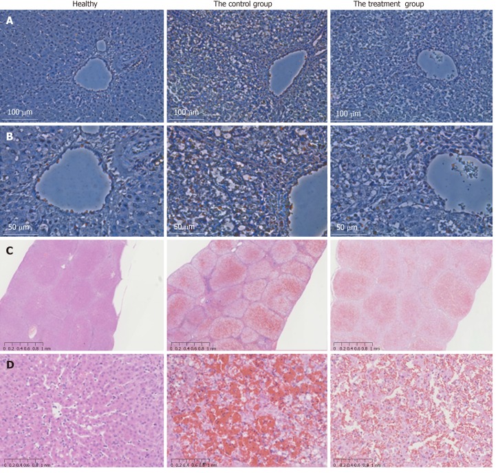
TUNEL assay and H&E staining of liver tissues after acute liver failure induction. A: Apoptotic hepatocytes were stained red. At 36 h after ALF induction, the number of apoptotic cells was significantly lower in the menstrual blood stem cell (MenSC) treatment group than in the control group (× 20); B: High magnification images were shown (× 40); C: The livers of the ALF pigs without MenSC treatment showed a typical ALF histology with extensive haemorrhage, hepatocyte necrosis and adipose degeneration at 48 h. In contrast, ALF pigs with MenSC transplantation showed increased numbers of remaining hepatocytes and hepatic parenchymal cells (× 2); D: High magnification images were shown (× 20). MenSC treatment alleviated the progression of liver injury. ALF: acute liver failure; MenSC: menstrual blood stem cell.

