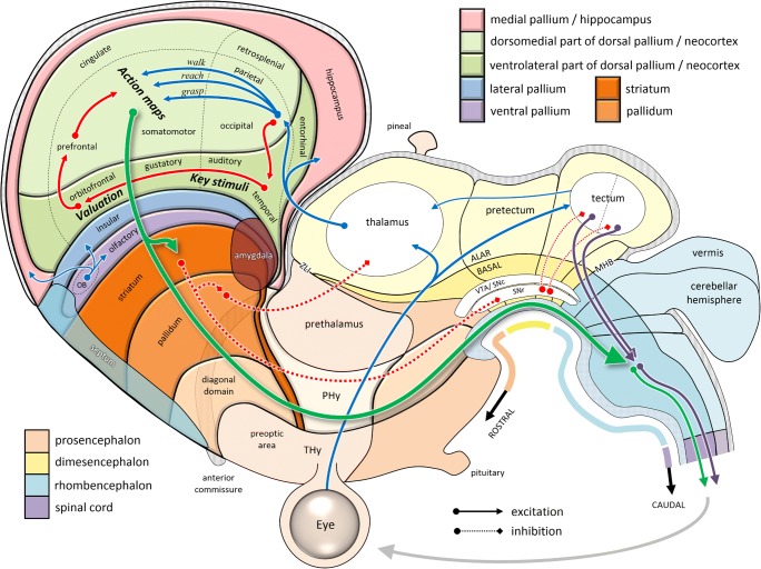
Fig. 7.
Schematic organization of the mammalian brain, based on Puelles et al. (2013). Here, the dorsal pallium (neocortex) has been divided into the spatially topographic (light) versus nontopographic (dark) neocortical sheets (Finlay & Uchiyama, 2015) and superimposed with labels based on the cortical flat map of Swanson (2000). Within the neocortical regions, blue arrows (see online color figure) indicate processes specifying potential actions, while red arrows indicate information related to their selection. Note the topological similarity of the tectal and telencephalic sensorimotor circuits to those shown in Fig. 6. OB, olfactory bulb; MHB, midbrain/hindbrain boundary; PHy, peduncular hypothalamus; SNc, substantia nigra compacta; SNr, substantia nigra reticulata; THy, terminal hypothalamus; VTA, ventral tegmental area; ZLI, zona limitans intrathalamica

