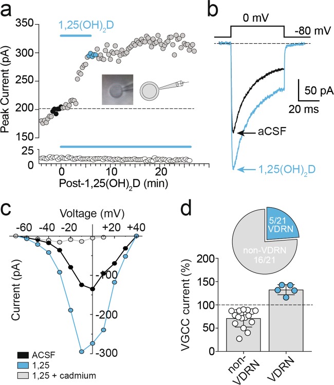
Fig. 2. 1,25(OH)2D enhanced high-voltage-activated (HVA) Ca2+ channel currents in a subset of PFC neurons.
a Baseline VGCC current amplitudes recorded in nucleated patch configuration (averaged in b, black trace) rapidly increased (averaged in b, blue trace) during bath application of 0.1 nM 1,25(OH)2D in a subset of PFC neurons (above), while the majority of recorded neurons were insensitive (below). Inset, representative and schematic examples of nucleated patch configuration following extraction from whole-cell configuration. c VGCC I–V recorded pre- (black) and post-1,25(OH)2D application (blue), abolished by cadmium (1 μM, grey). d 1,25(OH)2D evoked a mean increase in the recorded HVA Ca2+ current amplitudes of 33 ± 5% (blue circles, n = 5), while the majority of cells (open circles) demonstrated peak current amplitude rundown typical of L-VGCC channels (−29 ± 5%; n = 16). Data = Mean ± SEM

