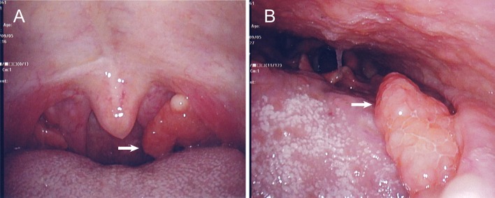
Fig. 1.
Gross images of lymphoid papillary hyperplasia of the left palatine tonsil. A polypoid lesion (arrowheads) is seen at the upper portion of the left tonsil (a) and demonstrates a cobblestone appearance when magnified by transnasal endoscopy (b). No polypoid lesion was seen in the right tonsil (a)

