Fig. 2.
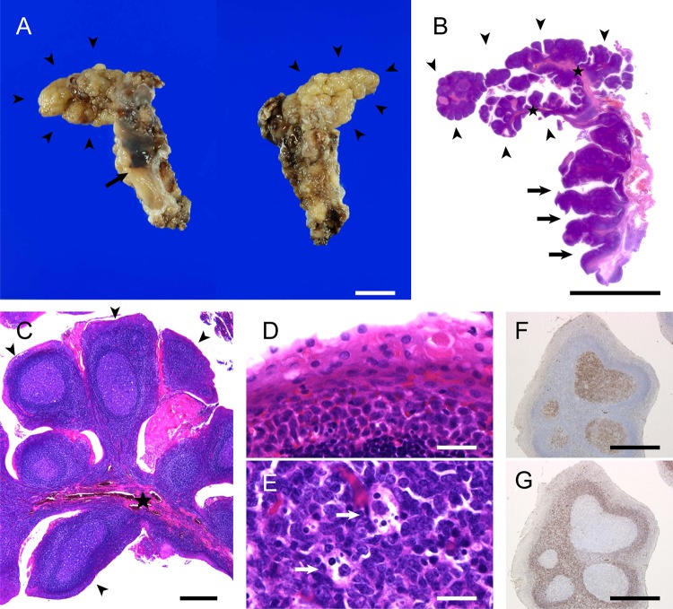
Macro- and microscopic findings of lymphoid papillary hyperplasia of the left palatine tonsil. a A polypoid lesion (arrowheads) with a cobblestone appearance is projected from the upper portion of the left palatine tonsil (left, anterior view; right, posterior view). No polypoid lesion is noted in the lower portion (arrow). b–e Microscopic appearance of hematoxylin-and-eosin-stained sections of the tumor. A longitudinally cut section of the resected tonsil demonstrates that the lesion consists of dome-shaped papillae with lymphoid stroma (arrowheads) protruding from fibrous central stalks (stars) (b–c). High magnification of the stratified squamous epithelium of the papillae demonstrates parakeratosis and no apparent atypia (d). Germinal centers have tangible body macrophages (e, arrows). f, g Immunohistochemically, the germinal centers are positive for CD10 (f) and negative for BCL2 (g). Scale bars in a and b: 1 cm; scale bars in c, f, and g: 500 µm; and scale bars in d and e: 20 µm

