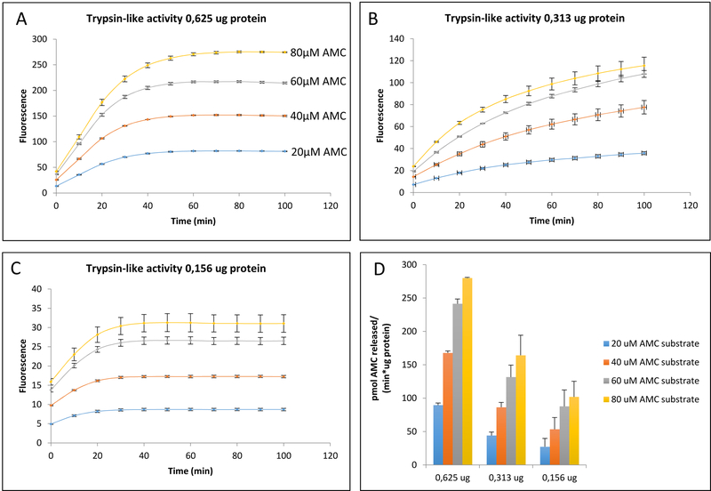
Figure 5: Testing Apparent Proteolytic Rates Across Varying Concentrations of AMC-conjugated Substrate and Cell Sample Protein.
A, B, C: proteolytic activity corresponding to different amounts of cell protein, each one tested against a variety of Boc-LRR-AMC (Proteasomal Trypsin-like) substrate concentrations. Proteasomal Trypsin-like apparent activity (fluorescence emission) initially increased with increasing concentrations of AMC-substrate, but the relationship was not linear. As AMC-conjugated substrate concentration was increased in increments of 20 uM, there was a gradual loss of consistent increases in fluorescence, with saturation at the higher concentrations that would negatively affect the accuracy of specific activity calculations. D: Specific activity corresponding to A, B, C. The apparent specific activity does not increase proportionally with increasing concentrations of AMC substrate. The results showed here are the mean ± SD of at least 3 independent trials.

