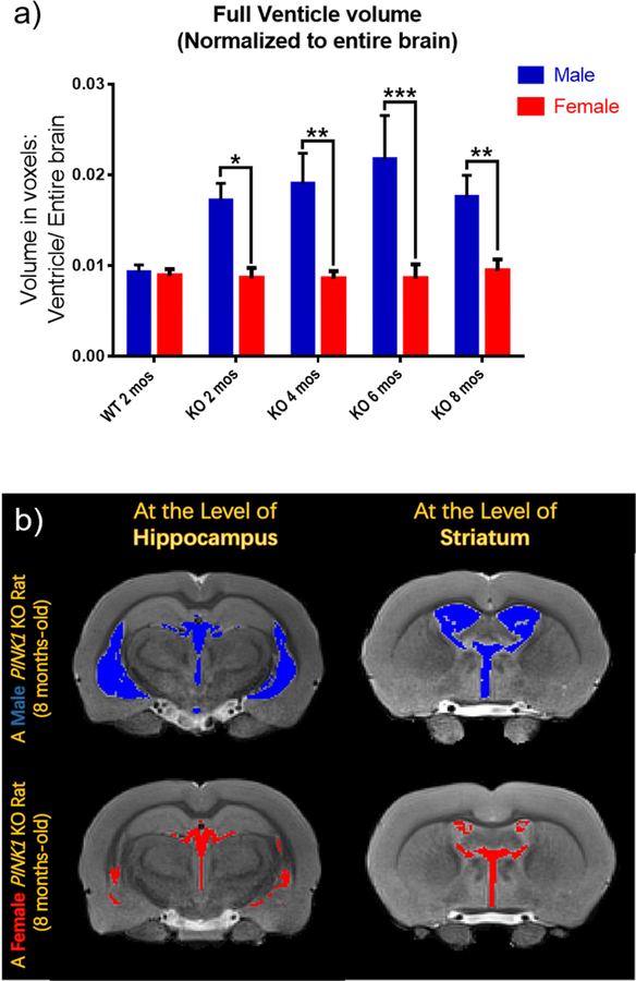
Fig. 2.
The ventricle sizes were measured from MRI images. The dimensions of the entire brain and all ventricles including two lateral ventricles, 3rd ventricle, and 4th ventricle were measured using the Waxholm Rat program after adjusting all the factors. The dimensions were acquired in voxels for each individual slice image of each brain. The values of each slice were added together to obtain the size of ventricles or the entire brain. The size of the ventricular system was normalized to the size of the entire brain. a) In the PINK1 KO rats, the male rats showed significantly larger sizes of ventricles in brain compared to those of females at the same ages. No differences were observed between males and females in the WT group. b) Representive images of brains from one male and one female from the group of KO rats at 8 mos-old. Brain images are presented at levels of both hippocampus and striatum. Ventricles of the male KO rat are marked in blue, while those of the female KO rat are marked in red. The images support the data showing in Fig. 2-a that male KO rats have larger ventricles than female KO rats.
*p<0.05, **p<0.01, ***p<0.001. For the data in Fig. 2-a, the numbers of individual rats were: male WT, N=3; female WT, N=3; male KO, N=6 for each age; female KO, N=6 for 2 and 8 mos and N=5 for 4 and 6 mos).

