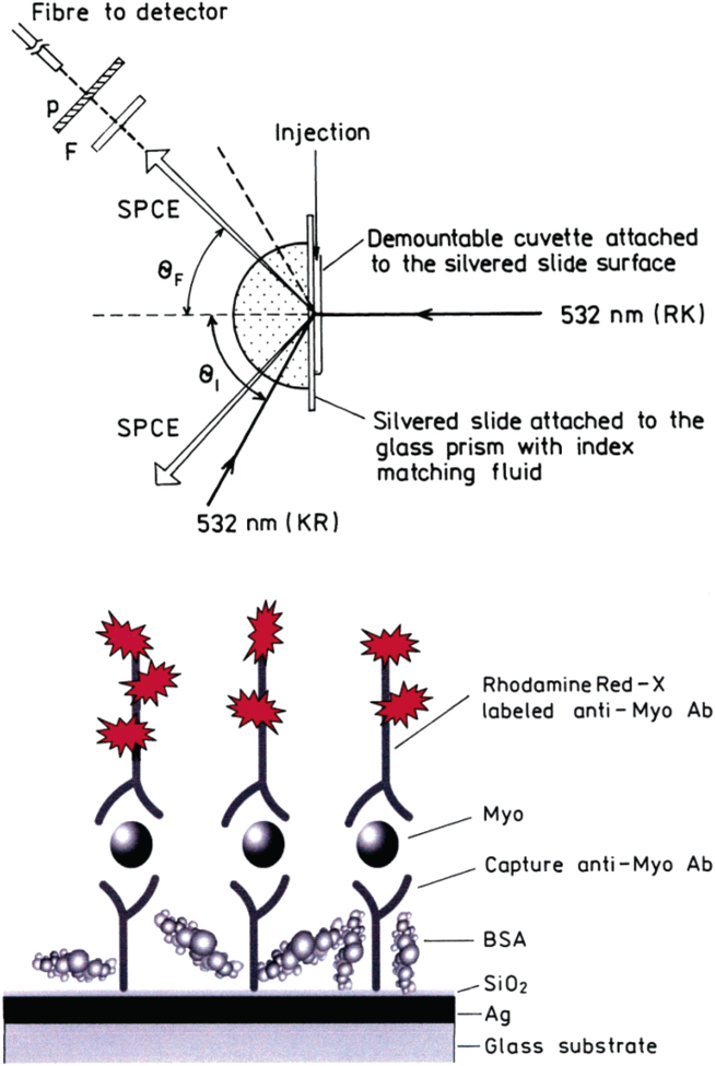
Figure 1.
Top: Experimental geometry for measurements of SPCE emission with RK and KR configurations. In RK configuration, the sample is excited directly by 532 nm and the emission out-couples through the prism in a hollow cone at the angle θF. In KR configuration, the excitation is provided by the evanescent field created by the incident light of 532 nm, which enters the system through the prism at the angle of θI. The fluorophores excited by this evanescent field couple to the surface plasmons, and the directional emission out-couples through the prism at the angle θF. The emission is collected by the fiber equipped with the filter (F) and polarizer (P). Bottom: Scheme of the myoglobin immunoassay (sandwich format) on a thin silver mirror slide surface. The drawing is not to the scale. The thickness of the silver layer was 50 nm and SiO2 protective layer 5 nm.

