Figure 1.

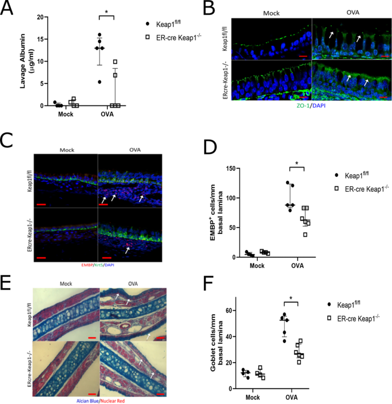
Keap1−/−; ER-cre mice demonstrate reduced barrier dysfunction in response to chronic Ova exposure. (A) Analysis of nasal lavage fluid for serum albumin (n= 4–6 per group). Mock is vehicle control. (B) Immunofluorescence for ZO-1 (green) demonstrates reduced barrier disruption. DAPI (blue). Images are at 100x magnification and scale bar is 10μm. (C) Immunofluorescence for eosinophil major basic protein (EMBP, red) and Keratin-5 (Krt5, green). White arrows indicate areas of increased eosinophil accumulation. Images are at 40x magnification and scale bar is 20μm. (D) Eosinophil counts per mm of basal lamina from imaging of the entire nasal septum (n= 4–6 per group). Goblet cell via Alcian blue stain (E-F) counts per mm of basal lamina (n= 4–6 per group). DAPI (blue). Images are at 20x magnification and scale bar is 50μm. * P<0.05, ** P<0.01, *** P<0.001, **** P<0.0001 Data are represented as median with interquartile ranges.
