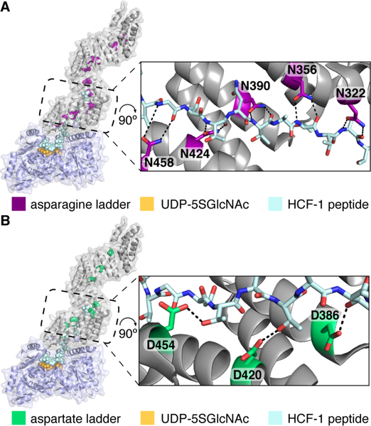
Figure 1.
Two conserved amino acid ladders line the OGT TPR lumen. A) Composite structure of human OGT complexed with a 26 residue peptide (light blue) was built by aligning overlapping residues from two structures (PDB codes 4N3B and 1W3B). Asparagine residues form a ladder, and the expanded view shows that five sequential asparagines closest to the active site make bidentate contacts to the bound peptide backbone. B) Composite structure as in A, but with TPR aspartates highlighted. Three sequential aspartates contact threonine sides chains of the bound peptide.

