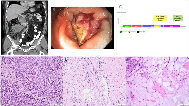
Figure 2.
Case #2 exhibiting treatment response to immune checkpoint blockade. At presentation, CT scan shows bulky peritoneal disease (A) and endoscopy revealed a lesion in the right colon (B). By Memorial Sloan Kettering-IMPACT, the biopsy sample of the tumor revealed three somatic, likely pathogenic MSH2 mutations (C). Resection of the tumor after 3 months of treatment with an anti-PD-L1 agent shows evidence of residual tumor with medullary features (D, similar to tumor in Fig. 1A), as well as evidence of tumor regression. The latter is seen in the form of fibro-inflammatory changes in some regions (E) and acellular mucin pools in other regions (F).

