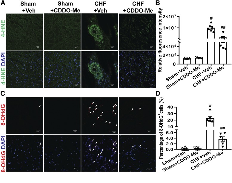
Fig. 7.
CDDO-Me decreases oxidative stress of the heart. Oxidative stress in the left ventricles of hearts was determined in sham and CHF rats (12 weeks post MI) that were treated with vehicle or CDDO-Me for an additional 2 weeks, respectively. (A) Representative confocal microscopic images of the left ventricle with 4-HNE staining. 4-HNE–positive is shown in green. (B) The relative fluorescence intensities were quantified by ImageJ software (NIH) (sham+Veh: n = 3; sham+CDDO-Me: n = 4; CHF+Veh and CHF+CDDO-Me: n = 6). #P < 0.0001 vs. sham+Veh; ##P < 0.0001 vs. CHF+Veh. (C) Representative confocal microscopic images of the left ventricle 8-OHdG staining. 8-OHdG–positive is shown in red. (D) The percentage of 8-OHdG+ cells was quantified (sham+Veh and sham+CDDO-Me: n = 4; CHF+Veh and CHF+CDDO-Me: n = 6). #P < 0.0001 vs. sham+Veh; ##P < 0.0001 vs. CHF+Veh. Nuclei are shown in blue (DAPI, 4′,6-diamidino-2-phenylindole). Original magnification, 400×. All images were processed with the same confocal settings.

