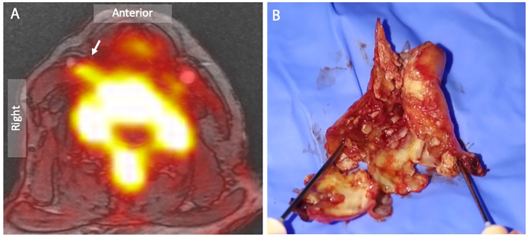
Figure 4.
18F-Sodium fluoride positron emission tomography and magnetic resonance angiography of a symptomatic right internal carotid artery lesion. (A) Combined 18F-Sodium fluoride positron emission tomography superimposed on MR angiogram localizes focal radiotracer uptake in the culprit right internal carotid artery plaque (arrow). (B) Surgical endarterectomy confirms a highly ulcerated lesion with positive remodelling and marked intimal irregularity

