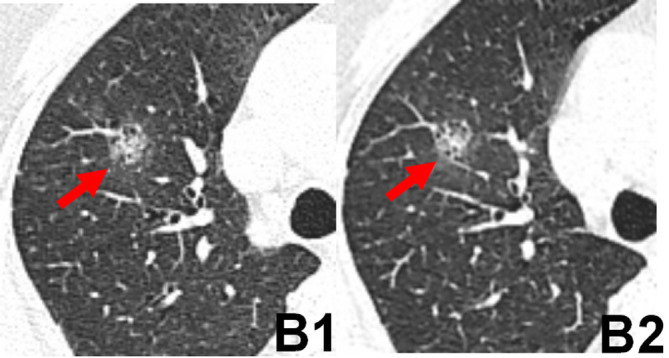
Figure 2. .

Transverse chest CT images of a 54-year-old male with a mixed-density pulmonary nodule in the middle lobe of the right lung. The edges of nodules (arrows) of images reconstructed with FBP in the standard-dose scan (B1) and 60% ASIR-V in the low-dose scan (B2) appeared blurred. The scores of the visibility of nodules of FBP image (B1) and 60% ASIR-V image (B2) were 3, 3, respectively. ASIR-V, adaptive statistical iterative reconstruction V; FBP, filtered back projection.
