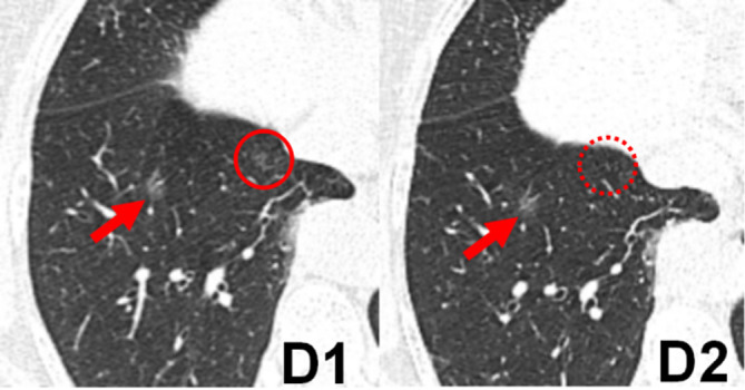
Figure 4. .

Transverse chest CT images of a 49-year-old female with a ground-glass pulmonary nodule in the lower lobe of the right lung. The edges of nodules (arrows) of images reconstructed with FBP in the standard-dose scan (D1) and 60% ASIR-V in the low-dose scan (D2) appeared blurred. The scores of the visibility of nodules of FBP image (D1) and 60% ASIR-V image (D2) were 3, 3, respectively. However, the other small ground-glass nodule was shown in the lower lobe of the right lung (D1, red solid circle), which was not clearly visible on the same level (D2, red dashed circle). ASIR-V, adaptive statistical iterative reconstruction V; FBP, filtered back projection.
