
Figure 1.
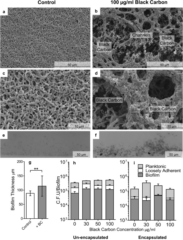
The effect of black carbon on S. pneumoniae biofilm structure. Biofilms of S. pneumoniae PR201 (a–h) and D39 (i) were cultured in the presence or absence of 30–100 μg/ml BC. Biofilms were imaged by Scanning Electron Microscopy (SEM) at increasing resolutions (a–d). Light microscopy was used to quantify biofilm thickness (e.g. n = 18). Images are representative of the entire biofilm structure. Viable bacterial cells were measured by sequential removal and quantification of planktonic, loosely‐adhered, and biofilm bacteria (h, i, n = 4). Error bars represent ± 1 SEM. Significance was determined by t‐tests (g) or ANOVA (h, i). **=p ≤ 0.01.