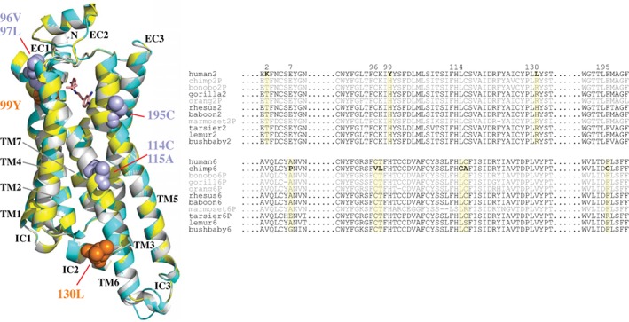
Figure 4.

Three‐dimensional‐structural model and partial sequence alignments for TAAR2 and TAAR6 proteins. (A) A 3D‐structural model of the human TAAR2 (yellow) and chimpanzee TAAR6 protein (cyan) superimposed with the Turkey β1‐adrenergic receptor (β1AR, gray). The ligand β1AR (dobutamine) is shown as a stick model. Positively selected sites are indicated in orange (human TAAR2) and dark cyan (chimpanzee TAAR6). Two positive selection sites (positions 2 and 7) are not shown due to the lack of a 3D protein model. (B) The partial sequence alignment of primate TAAR2 and TAAR6. The nine residues predicted to be under positive selection are shown in boldface (indicated by the yellow boxes). The pseudogenes are in gray
