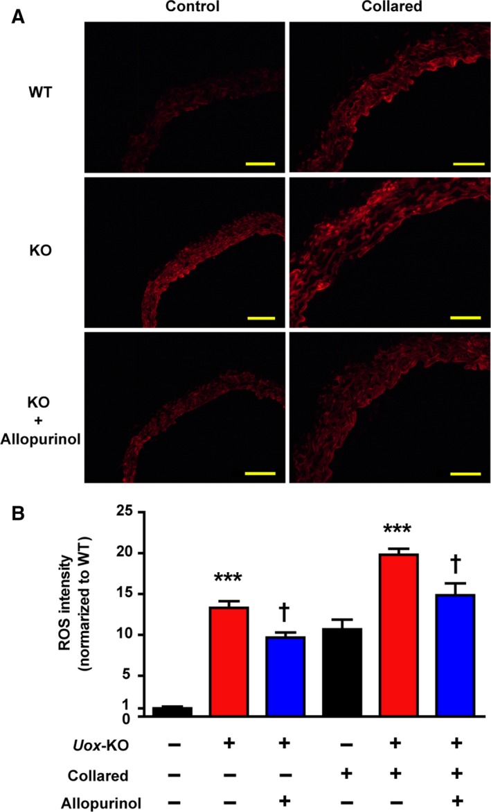
Figure 3.

HU elevates ROS in carotid artery in vivo. Carotid arteries were incubated with 2 μmol·L−1 DHE fluorescence probe for 30 min at 37 °C in the dark to measure ROS levels. (A) Representative images of fluorescent dye DHE staining from carotid artery of WT and Uox‐KO mice with or without collar placement are shown (males, n = 6). (B) Effects of 8‐week allopurinol treatment (100 mg·kg−1) on ROS were quantified. Scale bars = 50 μm. Error bars represent SEM. ***P < 0.001 vs WT control. † P < 0.05 vs untreated Uox‐KO mice (student's t test and one‐way analysis of variance followed by Newman–Keuls multiple comparison test).
