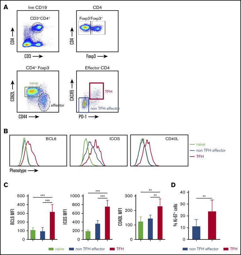
Figure 1.
FVIII immunization induces activated TFH cells in spleen. FVIIInull mice were given 5 rounds of weekly IV FVIII immunization injection. Five days after the last immunization, the mice were euthanized, and splenocytes and plasma were collected for analysis. Age-matched naive FVIIInull mice were used as controls. Anti-FVIII inhibitor titers were determined by chromogenic-based Bethesda assay. Splenocytes were stained for CD3, CD4, CD19, CD44, CD62L, Foxp3, CXCR5, PD-1, and Ki-67 and analyzed by flow cytometry. (A) Gating strategy for the identification of activated TFH cells. CD4+ Th cells were identified by excluding Foxp3+ CD4+ cells inside the singlet live CD19−CD3+CD4+ cell population. Effector CD4+ Th cells were identified by CD44 and CD62L expression (CD44+CD62L−). Effector CD4+ cells were analyzed for CXCR5 and PD-1 coexpression to identify TFH cells. (B) shows representative histograms and (C) shows quantification of BCL6, ICOS, and CD40L expression on naive, CXCR−PD-1− effector (non-TFH effector cells), and TFH cells (n = 6 mice per group). (D) Ki-67 cell cycling staining on CXCR5−PD-1− effector and TFH cells (n = 6 mice per group). **P < .01; ***P < .001.

