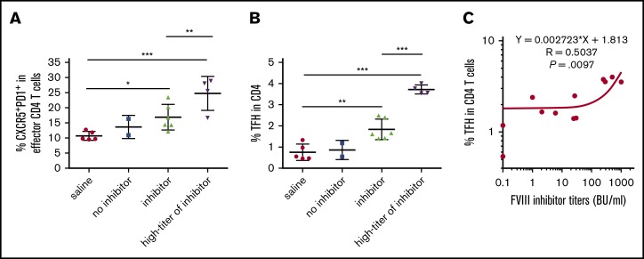
Figure 4.
Emergence of PD-1+CXCR5+TFH cells correlates with the development of FVIII inhibitors. FVIII and VWF double-knockout mice were given weekly IV FVIII immunizations. One week after the last immunization, the mice were euthanized. Splenocytes and plasma were collected for analysis. Age-matched saline-treated mice were used as a control. Plasma inhibitor titers were determined by Bethesda assay. Splenocytes were analyzed by flow cytometry. (A) Percentage of CXCR5+PD-1+ TFH cells among effector CD4-helper cells. Anti-FVIII inhibitor titers greater than 250 BU/mL were defined as the high-titer inhibitor group. (B) Percentage of activated TFH cells among whole CD4 T cells. Anti-FVIII inhibitor titers greater than 250 BU/mL were defined as the high-titer inhibitor group. (C) Dot-plot shows the correlation between the titer of FVIII inhibitor and the percentage of activated TFH cells among CD4 T cells. Statistical significance was analyzed by linear regression. The r and P values are indicated in the figure. *P < .05; **P < .01; ***P < .001.

