Figure 1.
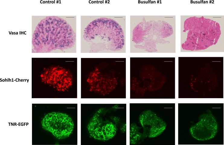
Imaging TNR (EGFP) expression following busulfan treatment of embryonic TNR/S1CF ovaries. E18.5 ovaries isolated from dams injected with vehicle (control) or busulfan at gestation day E11.5 are shown. Whole ovaries were used for confocal fluorescence microscopy to detect the Sohlh1-mCherry reporter or the TNR-EGFP reporter and were subsequently sectioned and processed for immunohistochemical detection of VASA protein. Original magnification, ×20; scale bars, 50 µm. Images from two representative animals are shown from n = 5 for each condition.

