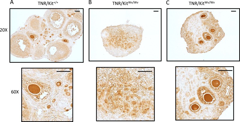
Figure 7.
TNR (EGFP) expression and localization in control and white-spotted mouse ovaries at PND19. (A) TNR/Kit+/+ ovary and (B) TNR/KitWv/Wv ovaries. PND19 ovaries were used for immunohistochemical detection of the EGFP Notch reporter. Original magnifications, ×20 and ×60; scale bars, 50 µm. These are representative images from n = 3 mice. The two TNR/KitWv/Wv ovaries shown illustrate one example without any apparent oocytes (left) and another with multiple remaining oocytes (right).

