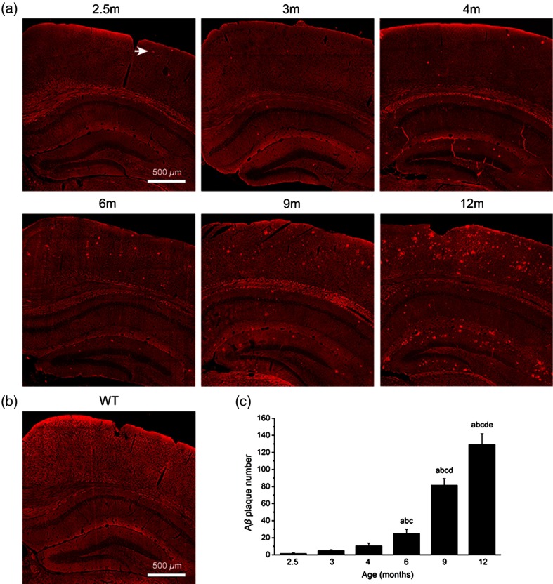
Fig. 4.
Progression of deposition with age. (a) TPEF images of extracellular plaques in CTX and HC from 2.5- to 12-month-old APP/PS1 mice. Arrow, plaque. (b) TPEF image from wild-type mouse. (c) Statistical analysis revealed that plaque number increases significantly with age. We performed ANOVA within groups. a, versus 2.5-month-old group. b, versus 3-month-old group. c, versus 4-month-old group. d, versus 6-month-old group. e, versus 9-month-old group. Each group has three mice.

