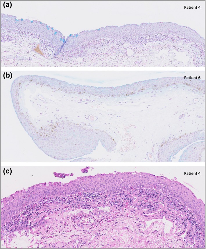
Figure 1.

Alcian blue‐stained histological sections of the inferior bulbar conjunctiva under light microscopy shows the presence of decreased goblet‐cell density in patients with AD treated with dupilumab (original magnification × 40). (a) Regions with no goblet cells (GCs) interspersed with smaller regions of normal GC density. (b) In patient 6 no GC was found in the conjunctival biopsy. (c) Haematoxylin and eosin stained histological sections of the inferior bulbar conjunctiva under light microscopy show the presence of a superficial inflammatory multicellular infiltrate in the conjunctival stroma consisting of mainly T cells and eosinophils, partially migrating into the conjunctival epithelium.
