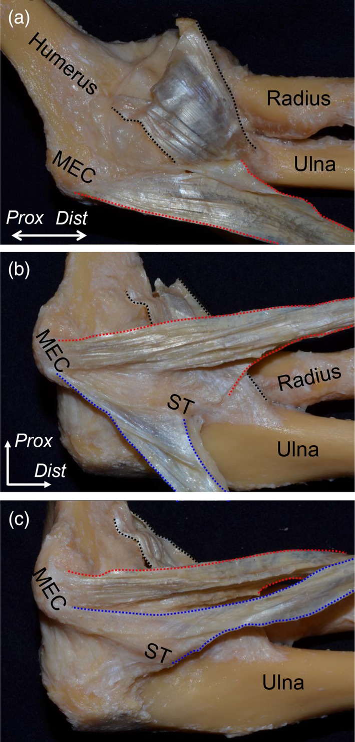
Figure 2.

Relationship between the TS and its surrounding structures. All muscular parts of the FPMs and nerves were removed. (A) The tendinous part of the brachialis muscle (black dotted area) and the anteromedial aspect of the TS between the PT and FDS muscles (red dotted area) of the left elbow are shown in the extended position. The brachialis tendon was mainly inserted into the coronoid process and partially connected to the base of the TS between the PT and FDS muscles at the sublime tubercle (ST) of the ulna. (B) In the flexion position of the elbow, the TS between the PT and FDS muscles was reflected anteriorly. It originated from the anterior slope of the medial epicondyle (MEC), distally extended to the anterior part of the ST, and transitioned into the deep aponeurosis of the FDS muscle over the humeroulnar joint. In addition, the TS between the FDS and FCU muscles was reflected posteriorly (blue dotted area). (C) The TS between the FDS and FCU muscles was reflected anteriorly. It originated from the posterior slope of the MEC, distally extended to the posterior part of the ST, and posteriorly transitioned into the deep aponeurosis of the FCU muscle over the humeroulnar joint. Dist, distal; Prox, proximal. [Color figure can be viewed at https://wileyonlinelibrary.com]
