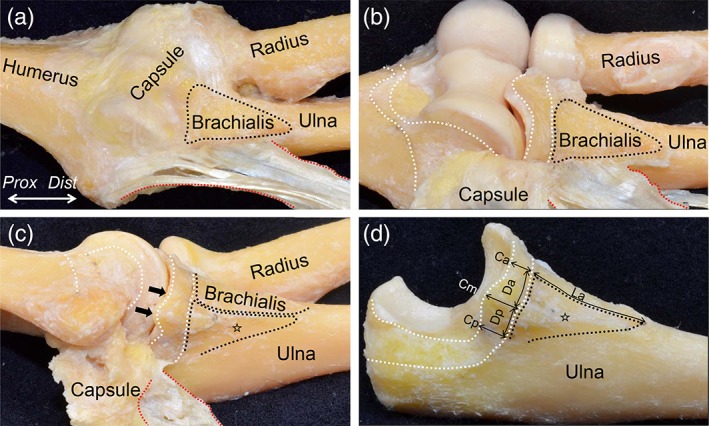
Figure 3.

Ulnar attachments of the tendinous complex and the capsule of the elbow joint. (A) The appearance after removing the brachialis tendon from Fig. 2 is shown. The ulnar insertion of the brachialis tendon (black dotted area) and the TS between the PT and FDS muscles (red dotted area) are shown. (B) The joint capsule was detached from the lateral to the medial margin of the brachialis insertion and medially reflected. The capsular attachments on the bones are shown as white dotted area. (C) Furthermore, we detached posteriorly the joint capsule and the tendinous complex containing the two TS and the deep FDS aponeurosis. The ulnar insertion of the tendinous complex is shown as the open star. The widths of the cartilage surface without the capsular attachment were too small to measure (black arrows). (D) Finally, the joint capsule and the tendinous complex were detached together from the ulna. The picture indicates measurements of the insertion of the tendinous complex and the attachment of the joint capsule on the ulna. Measured locations and data are shown in Table 1. Dist, distal; Prox, proximal. [Color figure can be viewed at https://wileyonlinelibrary.com]
