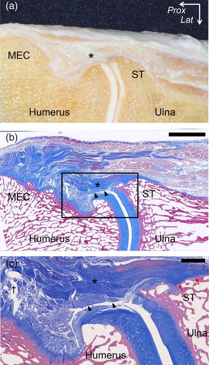
Figure 5.

Histological analysis of the oblique coronal section at the TS between the PT and FDS muscles. (A) The macroscopic view of the oblique coronal section at the level of the TS between the PT and FDS muscles is shown in Fig. 4A. (B) Masson's trichrome staining of the section is shown in A. The joint capsule (black arrowheads) was identified under the TS (asterisk). (C) Magnified image of the part is shown as black square in (B). Proximal to the humeroulnar joint, the capsule reflected to form a synovial cavity (black cross) with a distinct margin from the TS. Distal to the humeroulnar joint, the capsule intermingled with the TS and attached together to the sublime tubercle (ST) with a fibrocartilage (red arrows). Lat, lateral; Prox, proximal. Scale bar, 5 mm in B, 1 mm in C. [Color figure can be viewed at https://wileyonlinelibrary.com]
