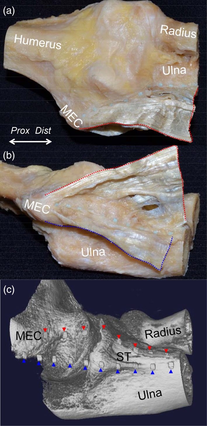
Figure 6.

Relationship between the TS and the bone morphology, showing the cadaveric sample of the elbow with the tendinous complex of the left elbow. (A) The radiopaque markers were set along the anterior base of the TS between the PT and FDS muscles (red dotted area). (B) The radiopaque markers were set along the anterior base of the TS between the FDS and FCU muscles (blue dotted area). (C) 3D imaging using micro‐CT of the sample with the radiopaque markers. The markers for the TS between the PT and FDS muscles are indicated as red arrowheads and those for the TS between the FDS and FCU muscles are indicated as blue arrowheads. MEC, medial epicondyle; ST, sublime tubercle; Dist, distal; Prox, proximal. [Color figure can be viewed at https://wileyonlinelibrary.com]
