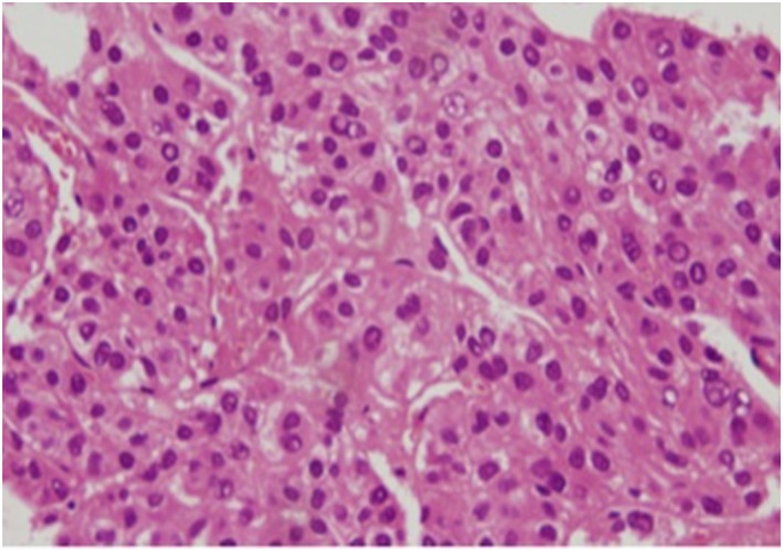
Figure 2.

Histopathological examination of liver biopsy sample before initiation of lenvatinib (hematoxylin–eosin staining, ×200) in a 68‐year‐old woman with unresectable advanced hepatocellular carcinoma (HCC) with portal vein invasion. The image shows moderately differentiated HCC tumor cells. [Color figure can be viewed at http://wileyonlinelibrary.com]
