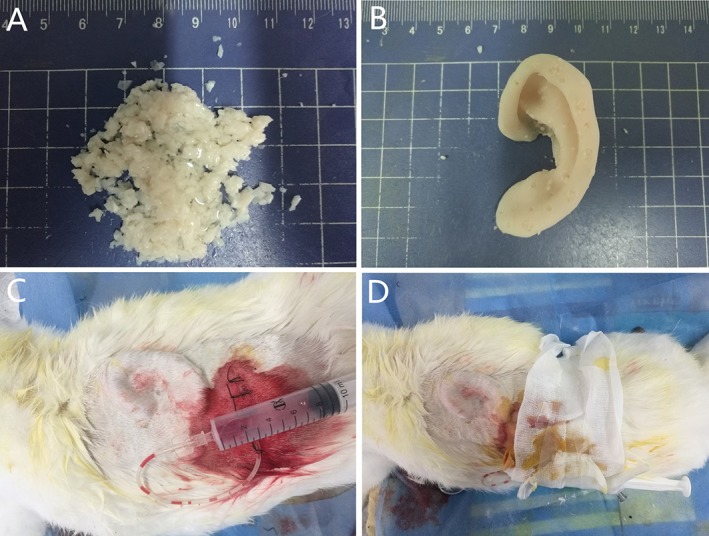
Figure 2.

Preparation of the graft and implantation in vivo. (A) Diced cartilage pieces were mixed with PRP. (B) The cartilage‐PRP mix was packed into the porous, hollow auricle mold. (C) The graft was embedded into the back of the rabbit with a negative pressure drainage device. (D) Gross view of the rabbit dorsum, showing the contour of the graft under the skin and dressing.PRP = platelet‐rich plasma. [Color figure can be viewed in the online issue, which is available at http://www.laryngoscope.com.]
