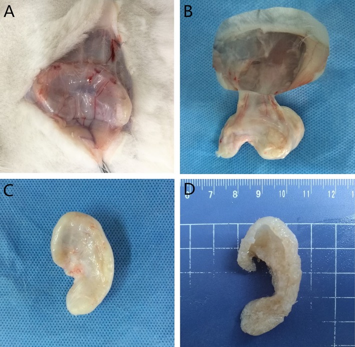
Figure 4.

The overall appearance of the auricle formed from diced cartilage mixed with PRP after 4 months. (A–B) Angiogenesis was observed from the periphery of the scaffold. (C) A fibrotic cyst had formed outside the scaffold. (D) The overall appearance of the auricle. [Color figure can be viewed in the online issue, which is available at http://www.laryngoscope.com.]
