Figure 8.

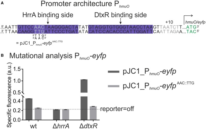
HrrA and DtxR cooperate to control hmuO expression in response to iron and heme availability. A. Promoter architecture of hmuO. The DtxR binding site was published previously (Wennerhold and Bott, 2006). The AAC::TTG mutation (grey letters) was shown to disrupt HrrA binding to PhmuO in vitro (Fig. S3). B. Mutational analysis of PhmuO. HrrA binding was abolished by introducing the AAC:TTG mutation into the PhmuO‐eyfp reporter. All strains were grown in BHI complex medium supplemented with 4 µM hemin as the ΔdtxR strain grows poorly in CGXII medium. After iron starvation overnight, the three strains (wild type, ΔhrrA and ΔdtxR) were inoculated in BHI and the specific fluorescence (eYFP‐fluorescence/backscatter) was recorded in 15 min intervals. The graph shows the specific fluorescence after 20 h. Full reporter outputs are shown in the supplementary Fig. S4 . [Colour figure can be viewed at wileyonlinelibrary.com]
