Figure 3.

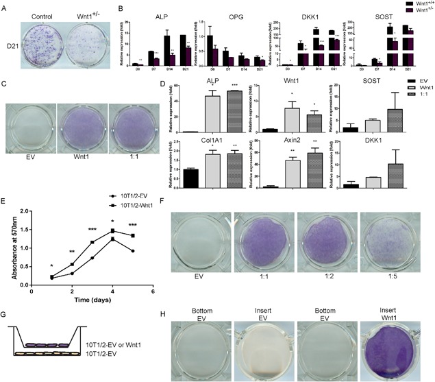
Mesenchymal cell‐derived Wnt1 stimulates osteoblast differentiation in a juxtacrine manner. (A, B) Calvarial osteoblasts from Wnt1+/+ and Wnt1+/– mice were induced to differentiate for indicated times. (A) ALP staining of control and Wnt1+/– calvarial osteoblast cultures. (B) mRNA expression levels of osteoblast‐related genes during calvarial cell differentiation measured by quantitative qRT‐PCR, was normalized to β‐actin expression and presented as a relative fold change to day 0 expression. (C) Mixed culture of Wnt1 overexpressing C3H10T1/2 cells with control EV‐transduced cells were stained for ALP at 7 days posttransduction. (D) mRNA expression of osteoblastic and Wnt target genes quantified by qRT‐PCR in cultures with 1:1 ratio of Wnt1 and control cells (EV). (E) Wnt1‐overexpressing C3H10T1/2 cells or control cells were seeded at density of 1500 cells/well. The CellTiter 96® Non‐radioactive Cell Proliferation Assay was used to detect cell viability at indicated time points. Results were presented as mean absorbance at 570 nm from triplicate assays. (F) Wnt1‐overexpressing C3H10T1/2 cells were growth‐arrested by mitomycin C and C3H10T1/2‐control (EV) cells were plated on top at different ratios keeping the total number constant. ALP staining was performed at D7. (G) Schematic of the method used to study Wnt1 effects mediated by secreted factors using cell culture well inserts. (H) ALP staining of cell culture wells (bottom) and inserts (insert) at D10. All cell culture experiments were repeated at least three independent times with at least three replicate wells. Staining and mRNA data are presented from one representative experiment as mean ± SD (n ≥ 3). B, D, and E: *p < 0.05, **p < 0.01, ***p < 0.001. Statistical analysis was performed by Student's t test. EV = empty virus.
