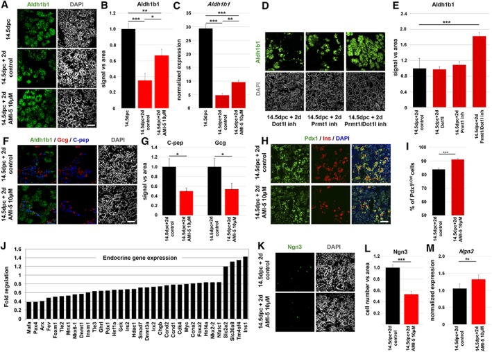
Figure 3.

AMI‐5 prolongs Aldh1b1 expression and delays endocrine differentiation in mouse embryo pancreatic explants. (A): Immunofluorescence analysis of pancreata at 14.5 dpc and after 2 days in ALI cultures (14.5 dpc + 2 days) shows that Aldh1b1 expression is reduced after 2 days in ALI culture of control pancreata whereas it remains higher in AMI‐5 treated pancreata. (B): Quantitation of relative Aldh1b1 fluorescence signal in 14.5 dpc pancreata and 14.5 dpc pancreata cultured in ALI for 2 days in the absence or presence of 10 μM AMI‐5 (n = 4). (C): qPCR analysis of Aldh1b1 expression in 14.5 dpc pancreata and 14.5 dpc pancreata cultured in ALI for 2 days in the absence or presence of 10 μM AMI‐5 (n = 6). (D): Immunofluorescence analysis of Aldh1b1 expression of 14.5 dpc pancreata after 2 days in ALI cultures in the presence of the Dot1l inhibitor EPZ004777 (10 μM) or the Prmt1 inhibitor C21 (10 μM) or both (10 μM each). (E): Relative quantification of the Aldh1b1 immunofluorescence signal shows significant upregulation in the presence of both inhibitors. (F): Immunofluorescence analysis of 14.5 dpc pancreata after 2 days in ALI cultures shows a reduction in the C‐pep and Gcg signal in pancreata treated with AMI‐5. (G): Relative quantitation of the C‐pep and Gcg fluorescence signal in 14.5 dpc pancreata cultured in ALI for 2 days in the absence or presence of 10 μM AMI‐5 (n = 4). (H): Immunofluorescence analysis of 14.5 dpc pancreata after 2 days in ALI cultures shows an increase of Pdx1LOW cells in pancreata treated with AMI‐5 and indicates the expression of ins in Pdx1HIGH cells. (I): Quantitation of the Pdx1LOW cells in relation to all Pdx1+ cells in 14.5 dpc pancreata cultured in ALI for 2 days in the absence or presence of 10 μM AMI‐5 (n = 3). (J): Fold regulation of endocrine markers at 14.5 + 2 days in ALI culture in the presence of 10 μM AMI‐5 in relation to untreated controls. Only significantly regulated genes are shown (padj ≤ 0.05). (K): Immunofluorescence analysis of 14.5 dpc pancreata after 2 days in ALI cultures shows a reduction in the number of Ngn3+ cells in pancreata treated with AMI‐5. (L): Relative quantitation of the Ngn3+ cells in 14.5 dpc pancreata cultured in ALI for 2 days in the absence or presence of 10 μM AMI‐5 (n = 4). (M): qPCR analysis of Ngn3 expression in 14.5 dpc pancreata cultured in ALI for 2 days in the absence or presence of 10 μM AMI‐5 (n = 3). *, p < .05; **, p < .01; ***, p < .001, error bars show SEM. Scale bar: 50 μm. Abbreviation: ALI, air to liquid interface.
