
Figure 5.
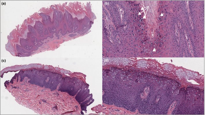
Histological representative cases of classical cutaneous viral warts. (a) Verruca vulgaris: haematoxylin and eosin (H&E) low‐power view (original magnification × 50) with architectural characteristic inturning of the elongated rete ridges, epidermal hyperplasia, papillomatosis, hypergranulosis, hyperkeratosis and columns of parakeratosis. (b) Verruca vulgaris: H&E, detail view (× 200). Note koilocytes (arrowheads) and coarse granuloma (arrows) mostly in the top layers (stratum granulosum). (c) H&E low‐power view (× 50) of verruca plana with epidermal hyperplasia, hypergranulosis, hyperkeratosis and koilocytes in the middle and upper layers. (d) Verruca plana: H&E, detail view (× 100). Note the absence of papillomatosis, parakeratosis and coarse granuloma.