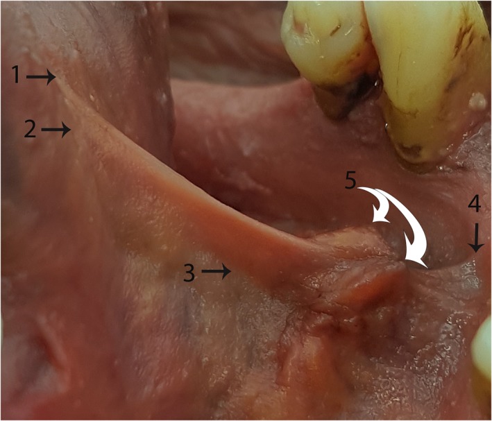
Figure 5.

“Surface anatomy” of the lingual frenulum—Example 1 Tongue elevated to create tension in the floor of mouth fascia, raising the fold of the frenulum. (1) Highest point of midline mucosal attachment to ventral tongue. (2) Highest point of midline floor of mouth fascia attachment to ventral tongue. (3) Genioglossus—drawn into base of lingual frenulum (suspended from floor of mouth fascia). (4) Highest point of midline fascial attachment on the inner surface of mandible. (5) (White arrows): Submandibular duct openings—suspended from floor of mouth fascia. [Color figure can be viewed at http://wileyonlinelibrary.com]
