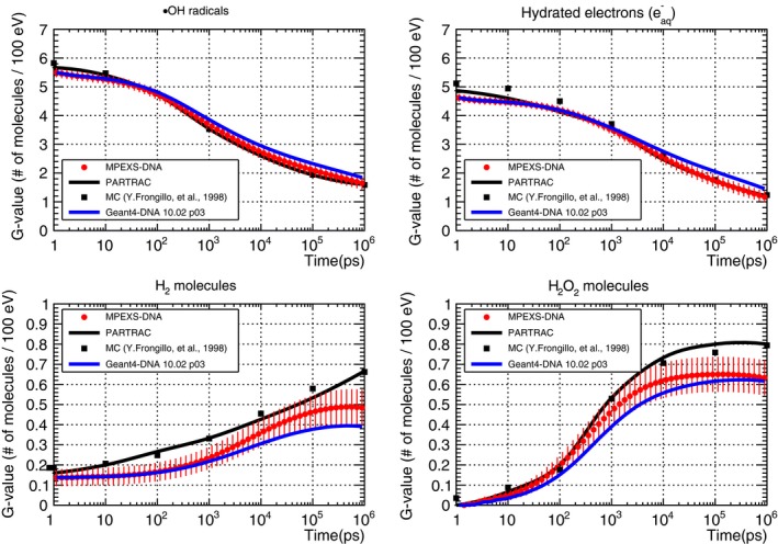
Figure 9.

G‐value time profile for radicals (top left), hydrated electrons (top right), H2 (bottom left), and H2O2 molecules (bottom right) induced by 5 MeV protons. MPEXS‐DNA is represented by filled red circles with vertical bars representing the standard deviation. Solid blue and black lines are Geant4‐DNA 10.02 p03 and PARTRAC23 results, respectively. Filled squares are Monte Carlo simulation results by Frongillo et al.44
