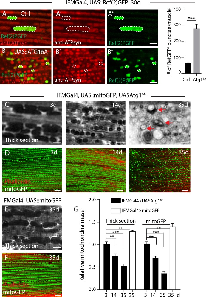
Fig 2. Age dependent accumulation of ubiquitinated mitochondria proteins are recycled by Atg1-related autophagy.
A-B, IFMs of 30-day old flies expressing Ref(2)PGFP (in green) was labeled with anti-ATP syn(red) for mitochondria. Typical mitochondria are circled in dashed lines. Genotypes: A, IFMGal4; UAS::Ref(2)PGFP, and B, IFMGal4; UASAtg16A, UAS::Ref(2)PGFP. C, Thick section of muscles from different time points, “vacuolation” of mitochondria are denoted as arrows. Mitochondria are densely stained by Toluidine blue in C and E. D, Mitochondrial mass indicated by matrix targeted mitoGFP (green) from different time points. Muscle fibers were stained with Phalloidin in red. Genotype in C-D, IFMGal4; UAS::mitoGFP, UASAtg16A. E, Thick section of muscles from 35-d old control flies. F, Mitochondrial mass indicated by matrix targeted mitoGFP (green) in control flies, and Phalloidin stained muscle fibers in red. Genotype in E and F: IFMGal4; UAS::mitoGFP. G, Relative mitochondria mass in C-F was quantified. At least 20 muscles from 6 thoraxes of each condition were analyzed. t-Test was performed for statistics, p value for S.E.M, **: p<0.01, ***: p<0.001.

