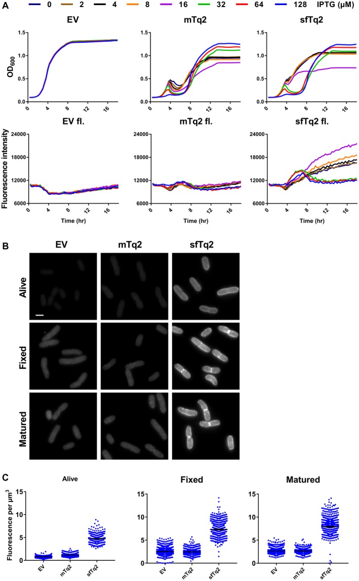
Figure 3.

Superfolder mTurquoise2 fluoresces in the periplasm. A. Plate reader growth data in rich medium at 37°C show that the periplasmic sfTq2 expression is less toxic at high induction than the original mTq2. Plate reader fluorescence measurements confirm that periplasmic sfTq2 gives fluorescent signals while mTq2 does not. B. Fluorescence microscopy of living cells expressing an empty vector control, mTq2‐PBP5 or sfTq2‐PBP5, reveals fluorescence only from the sfTq2 fusion. Fixation of the same cells does not result in a decreased sfTq2 signal. Overnight maturation of the fixed cells at RT to allow for possible chromophore (re)formation (indicated as ‘matured’) did not result in an increase in fluorescence for periplasmic mTq2 suggesting it had not folded properly while sfTq2 showed a strong periplasmic signal. All photographs are shown with the same grey values (100–4000) for comparison and the scale bar represents 2 µm. C. Quantification of the control, mTq2 and sfTq2 cultures for the living, fixed and matured cells shows no difference between the empty vector control and the mTq2 cells. The sfTq2 cells showed a small maturation effect. The error bars at the mean represent the 95% confidence interval. The number of cells measured were: Alive) EV = 1018, mTq2 = 650 and sfTq2 = 571. Fixed) EV = 1176, mTq2 = 1046, sfTq2 = 884. Fixed and matured) EV = 1142, mTq2 = 931, sfTq2 = 988.
