. 2019 Oct 30;8:e50051. doi: 10.7554/eLife.50051

Figure 4. Sphingomyelin depletion potentiates hedgehog signaling at the level of Smoothened.

(A) HH signaling triggered by HiSHH (25 nM) in the presence or absence of Vismodegib (2.5 µM) in NIH/3T3 cells treated with myriocin. (B) Human SMO in complex with cholesterol (PDB 5L7D) highlighting two residues in the CRD (D95 and Y130) critical for cholesterol binding and a residue (D473) in the transmembrane domain (TMD) critical for binding to the agonist SAG. Numbering for mouse SMO, used in our studies, is denoted in parenthesis. (C) HH signaling triggered by myriocin alone in Smo-/- MEFs stably expressing the indicated variants of mouse SMO. A control experiment (right) shows that the SMO variants respond appropriately to either SAG (100 nM) or SHH (50 nM), demonstrating protein integrity. Note that the D477G and D99A/Y134F mutations abrogate responses to SAG and SHH, respectively (Luchetti et al., 2016). In (A) and (C), bars denote the mean value derived from the measurements shown (n = 4 for A, n = 2–4 in C). Statistical significance (p-value=0.0286 for both A and C) was determined by the Mann-Whitney test.

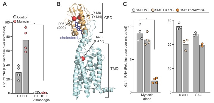
Figure 4.

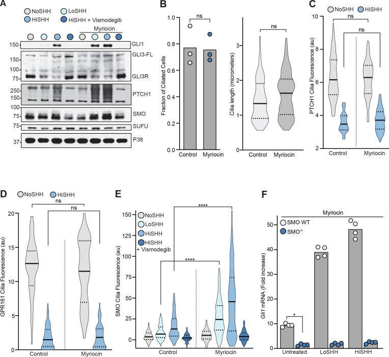
Figure 4—figure supplement 1. The effect of myriocin treatment on the abundances of hedgehog pathway components and on ciliary protein trafficking.

Figure 4—figure supplement 1.

(A) Western Blot showing levels of HH signaling proteins (SMO, SUFU and GLI3-FL/GLI3R) and HH target genes (GLI1 and PTCH1) after treatment with myriocin and either LoSHH (2.5 nM), HiSHH (25 nM) or HiSHH with Vismodegib (2.5 µM). (B) Ciliation frequency (left) of cells after myriocin treatment, calculated as the number of cilia over the number of nuclei. Each point represents the ciliation frequency (derived from >30 cells) in a different imaging field. The cilia length distribution (from 100 cells) is shown on the right. (C and D) Violin plots showing the lack of an effect of myriocin on levels of PTCH1 (C, n > 50 cilia per condition) or GPR161-YFP (D, n > 35 cilia per condition) at cilia by quantitative fluorescence microscopy in the absence or presence of HiSHH (25 nM). (E) Violin plots showing the SHH-induced ciliary accumulation of endogenous SMO in the presence and absence of myriocin (n > 100 cilia per condition, SHH concentrations as in A). (F) HH signaling triggered by myriocin alone or myriocin in combination with LoSHH (5 nM) or HiSHH (50 nM) in Smo-/- MEFs or Smo-/- MEFs stably expressing wild-type SMO. Bars denote the mean value derived from the four individual measurements shown. Statistical significance was determined by the Mann-Whitney test (B–F); p-values are: (B) p-value>0.9999 for fraction of ciliated cells and p-value=0.0603 for cilia length, (C) p-value=0.7581 for NoSHH and p-value=0.7801 for HiSHH, (D) p-value=0.5469 for NoSHH and p-value=0.5604 for HiSHH, (E) p-value<0.0001 (both comparisons), and (F) p-value=0.0286.