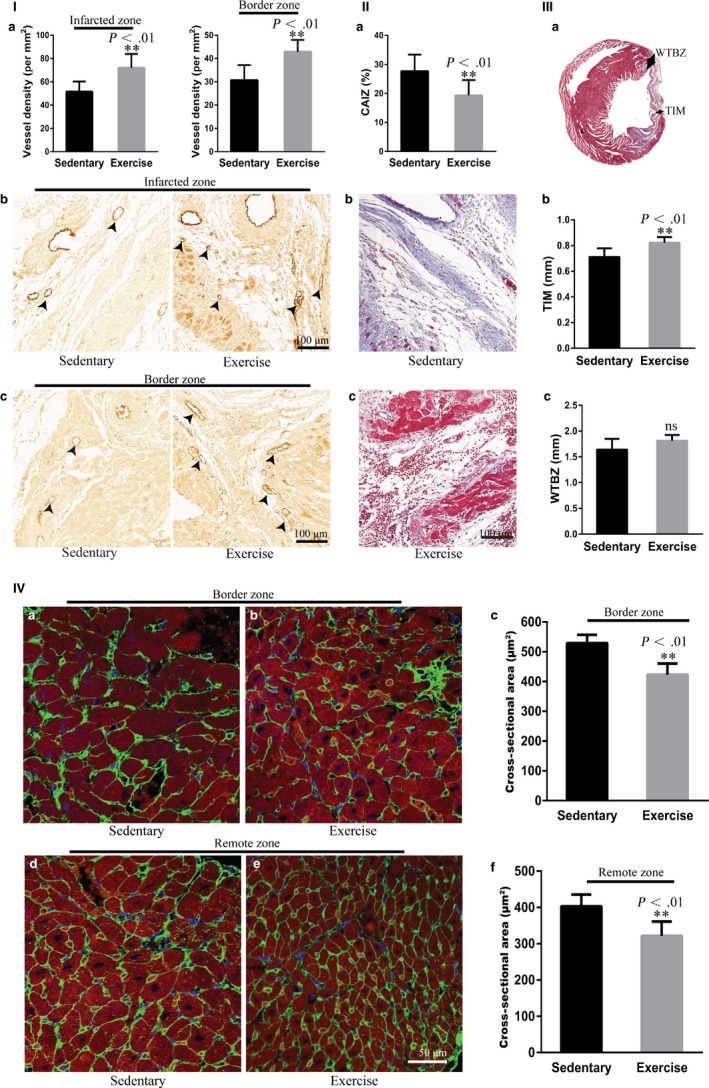
Figure 2.

Early moderate exercise improves angiogenesis, fibrosis and ventricular remodelling in myocardial infarct (MI). (I) Immunohistochemical staining for vWF showed that blood vessel density in the infarct zone and border zone in early moderate exercise hearts was significantly higher than those in sedentary hearts. (a) Semiquantification of (b) and (c). (b) Infarct zone. (c) Border zone. n = 6‐8. (II) Masson's trichrome staining documented that the collagen area of the infarct zone in early moderate exercise hearts was significantly smaller than that in sedentary hearts. (a) Semiquantification of (b) and (c). (b) Sedentary heart. (c) Early moderate exercise heart. n = 9 per group. (III) Masson's trichrome staining revealed that the TIM in early moderate exercise hearts was significantly larger than that in sedentary hearts, whereas the difference in the WTBZ between early moderate exercise hearts and sedentary hearts was not significant. (a) Schematics of TIM and WTBZ. (b) Semiquantification of TIM. (c) Semiquantification of WTBZ. n = 6‐9 per group. (IV) WAG immunofluorescence staining demonstrated that the cardiomyocyte cross‐sectional area of both the border zone and the remote zone in early moderate exercise hearts was significantly smaller than those in sedentary hearts. (a) Border zone of the sedentary heart. (b) Border zone of the early moderate exercise heart. (c) Semiquantification of (a) and (b). (d) Remote zone of the sedentary heart. (e) Remote zone of the early moderate exercise heart. (f) Semiquantification of (d) and (e). n = 5 per group. The animals were trained on early moderate exercise for two weeks beginning one day after MI
