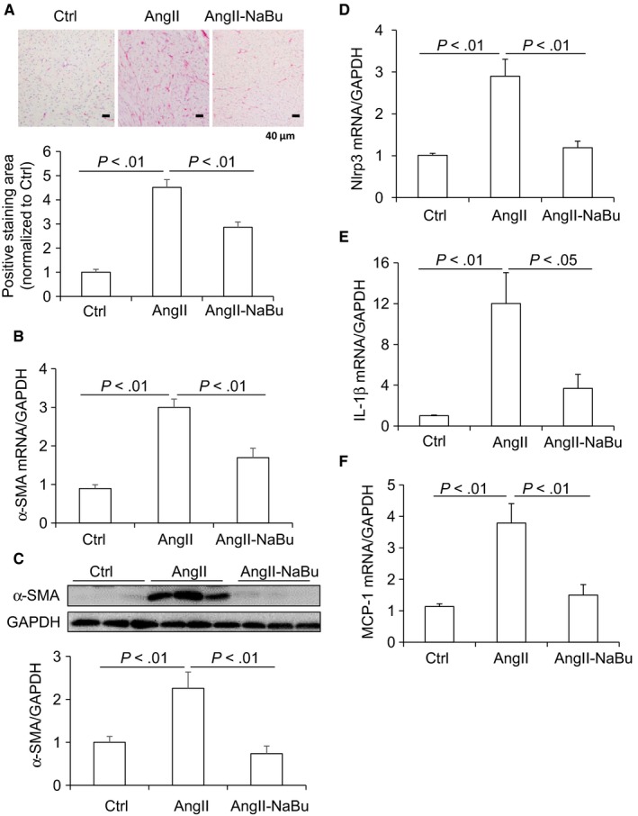
Figure 3.

NaBu decreased Ang II‐induced cardiac fibrosis and inflammatory factors. A, representative photomicrographs showing Picro‐sirius red staining of collagens in heart (red colour) and calculated percentage of positively stained area. B, mRNA level of α‐SMA in hearts. C, Representative immunoblots of α‐SMA protein levels in hearts and summarized intensities of blots. D‐F, mRNA levels of inflammatory factors Nlrp3, IL‐1β and MCP‐1. N = 6 per group
