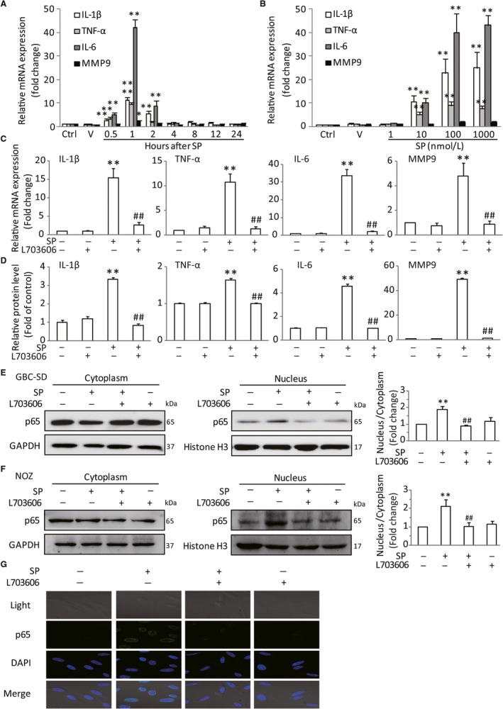
Figure 3.

Activation of NK‐1R could promote NF‐κB activation in vitro. GBC‐SD cells were stimulated by SP with different dose for the indicated time. The mRNA production of tumor‐related cytokines IL‐6, IL‐1β, TNF‐α and MMP9 were determined by real‐time PCR, and the expression levels were normalized to β‐actin (A and B). After pretreated with L703606, the cells were stimulated with SP, the mRNA expression and protein levels in GBC‐SD cells were analysied (C and D). The NF‐κB p65 nuclear translocation in GBC‐SD and NOZ cells were assessed by western blot. Histone H3 and GAPDH were used as nuclear and cytoplasmic markers (E and F). Immunofluorescence was used to analyze NF‐κB p65 nuclear translocation (scale bar, 20 μm) (G). Data are presented as means ± SEM (n = 6). Statistical analysis was performed using one‐way ANOVA coupled with a post hoc test. Significant differences were indicated as *P < 0.05 versus Ctrl, **P < 0.01 versus Ctrl, ## P < 0.01 versus SP
