Figure 3.

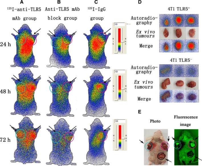
Whole‐body phosphor‐autoradiography and fluorescence imaging. Representative images were obtained at 24, 48 and 72 h post‐injection of 125I‐anti‐TLR5 mAb and showed apparently radioactivities in TLR5+ 4T1 tumour (A). Representative images showed no significant radioactivity accumulation TLR5+ 4T1 tumour at 24, 48 and 72 h post‐injection of 125I‐antiTLR5 mAb in block group (30 min prior to injection of 125I‐anti‐TLR5 mAb, 100 μg anti‐TLR5 mAb in 100 μL was injected through the tail vein (B). Representative images were obtained at 24, 48 and 72 h post‐injection of 125I‐IgG (C). Representative images of isolated tumours (isolated from model mice in Figure 3A at 72 h) (D). Representative fluorescence image of 4T1 tumour‐bearing mouse model (E).For A, B, C and E, the arrow on the left referred to the TLR5‐ tumour (blue circle), and the right is TLR5+ tumour (pink circle). For E, the bottom arrow referred to the TLR5+ tumour (without lentivirus transfection) (black circle). The data are from three independent experiments
